The Brain: Memories and Emotions
Have you ever wondered how your brain really functions—your thoughts, your memories and how we, humble humans, can achieve the feats we do? Well, wonder no longer, as this article will unveil how memories and emotions are created through phenomena such as active potentials and more

Have you ever wondered how your brain really functions—your thoughts, your memories and how we, humble humans, can achieve the feats we do?
Well, wonder no longer, as in this article I will show you how memories and emotions are created through phenomena such as active potentials and synaptic strengthening. I will then go on to describe cases of personality changes after neurological activity has been disrupted, most commonly through neurosurgery, and explain how this can occur. So read on, dear reader, and prepare to have your mind blown (excuse my pun).
Getting Fired
Section One
"In the beginning there was light."
The formation of memories begins due to an impulse from external stimuli, such as light, which is taken by the eye and converted into electrical signals via transduction (in a sensory receptor).
Suppose this signal is strong enough to raise the membrane potential (the voltage difference across a neuron membrane caused by uneven ion distribution on each side) past the activation threshold of -55 mV (a negative voltage as the inside has a negative charge relative to the outside), from the resting potential of -70 mV (in other words, the membrane potential of a neuron that is not active and waiting for a stimulus to activate it - the signal raises the charge inside the neuron).
- In that case, an action potential is triggered, which is a rapid sequence of changes to the voltage across the membrane of a neuron due to sodium and potassium ion channels as the positive sodium ions move into the cell via the sodium channel, creating an increasingly positive voltage inside the neuron cell (stages A-B on the graph in Figure 1).
Once, however, overshooting has occurred (B), the sodium channel shuts, and the voltage-gated potassium channels open (at about +30 mv), allowing potassium to move out of the cell until the resting potential of -70 mv is reached (C), and the neuron stops being activated as the activation threshold is no longer surpassed.
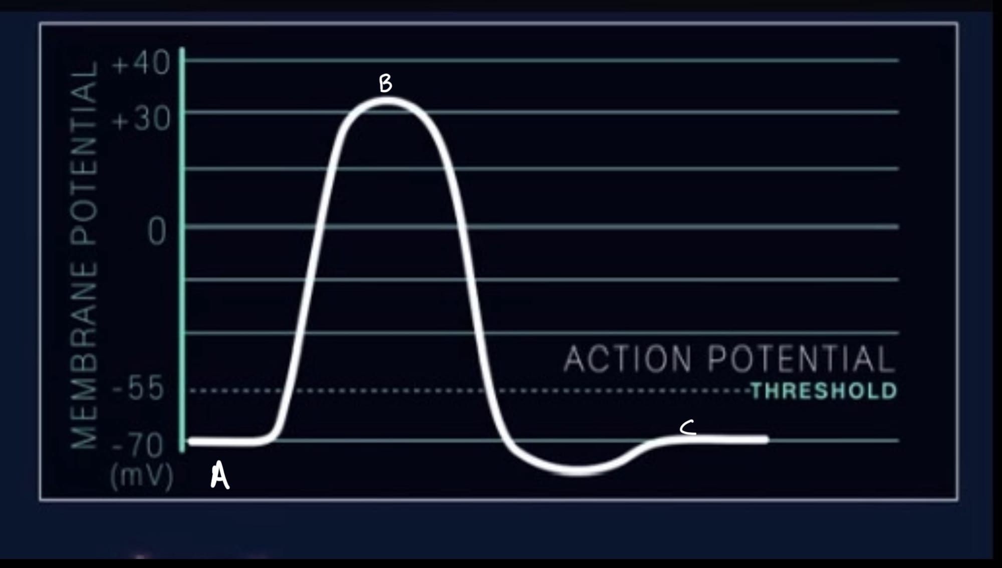
This entire cycle is also known as the “firing” of a neuron, where the membrane potential spikes and then goes back to normal.
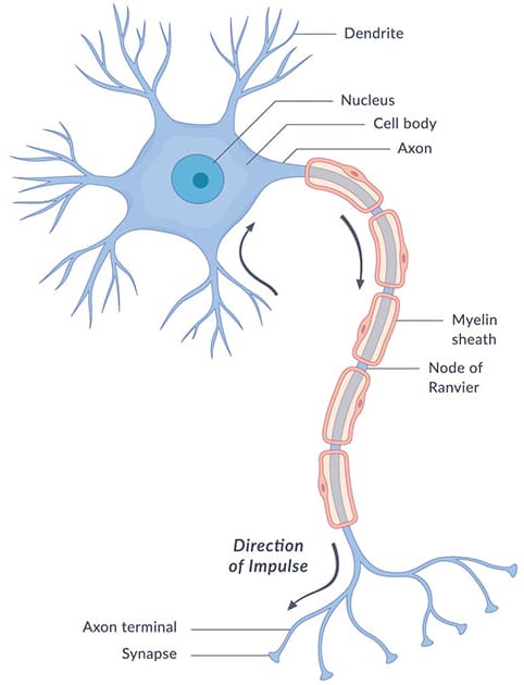
This will happen to the membrane all the way down the axon until the axon terminal, which transmits the signal to the dendrites of the next nerve cell. (If you are unsure on the anatomy of the nerve and completely clueless about what a dendrite is, take a gander at the diagram below)
Memories
Section Two
Via the concept of Hebbian Learning (which explains how neurons adapt and form stronger connections through repeated use), information is stored when synapses that connect to a group of neurones become more (or less) able to generate an action potential. When a neurotransmitter (which are chemicals to transmit the signal across nerve cells e.g. Glutamate) is released repeatedly with the firing of neurones in the nerve, an increase in the strength of the synapse is generated, due to the incorporation of more AMPA receptors (more receptors mean a quicker and stronger connection between neurones).
This strengthening of the synapse is through synaptic plasticity, particularly long-term potentiation (LTP), which is where the repeated activation of synapses leads to increased sensitivity of those synapses, causing the addition of more receptors (e.g., NMDA and AMPA receptors) on the (postsynaptic) neuron, making it easier for future activation.
This change is what is needed to form memories.
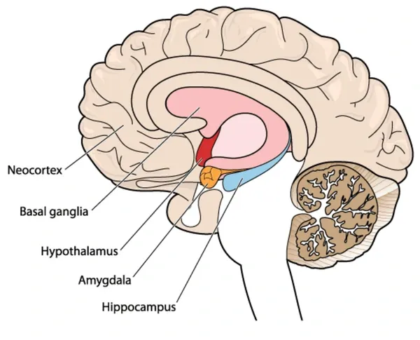
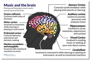
The Hippocampus (see diagram for anatomy of the brain) then stores this newly encoded memory and transfers it via white matter (a collections of axons) to the Neocortex (particularly the two temporal lobes in the lower end of the neocortex) for long-term storage (if the memory is factual rather than episodic; episodic memories are stored in the medial temporal lobes - a small area in each temporal lobe near the middle of the brain).
This is theorised to be due to the replay of neural activity during sleep, where the patterns of firing are reactivated between the Hippocampus and Neocortex, strengthening the synapses in this region and ensuring that the memory is integrated into the cortical networks.
To retrieve these memories, the neurons in the Prefrontal Cortex and Hippocampus are fired, reactivating the stored patterns of the Neocortex and allowing the memory to be reconstructed into conscious awareness.
Emotions and the Limbic System
Section Three
Emotions and memories are directly linked, with many of our strong memories being due to emotions and vice versa. This connection between the two is mainly due to the chemical fluctuations in the brain from emotions, which affect the brain's ability to store memories by “attaching” emotions to a certain memory.
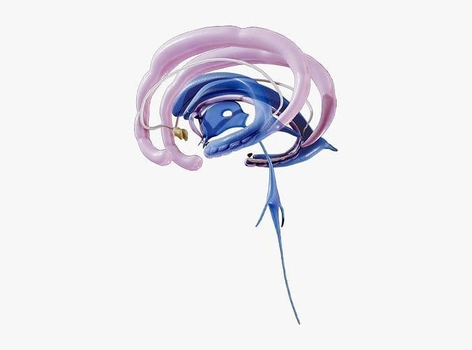
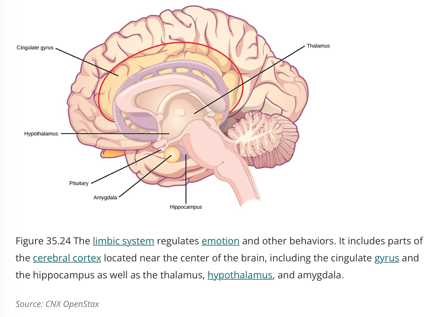
Emotions cause a series of cognitive, physiological, and behavioral responses within the brain. The key to emotion is predominantly from the limbic system, which is a collection of structures within the brain that collaborate, the key structures being the Hypothalamus, the Amygdala, the Thalamus and the Hippocampus, which all sit above the brain stem as can be seen in below.
The Thalamus relays impulses from stimuli to appropriate brain centers, where they will trigger the firing of neurones in that structure.
For example, the stimulation of the amygdala will result in emotions such as anger, fear, and anxiety. Destroying (which is the term for "unstimulating" neurones) the amygdala will result in mellowing. This was first noted by Dr. Kluver and Bucy with bilateral destruction, known as Kluver-Bucy syndrome, resulting in hyperorality, hypersexuality, and disinhibited behaviour (meaning you will ignore social conventions and act very irrationally, not considering risks or danger). In some individuals, alcohol (particularly in alcoholics) can excessively stimulate the amygdala to a large degree, leading to extreme anger and a heightened state of emotions.
The amygdala is quite sensitive towards emotionally stimulating stimuli (such as threats and rewards). When this information is perceived as emotionally important by the Thalamus, the amygdala's neurons become active and interact through synaptic transmission (via neurotransmitters such as glutamate) to other brain regions. For example, if there is a threat, the amygdala will fire, resulting in the release of glutamate to activate the hypothalamus, stimulating the “fight or flight response.". This triggers the autonomous nervous system to release hormones, and increase heart rate and sweat, correlating with the emotion of fear or excitement.
The amygdala's activity is modulated by the prefrontal cortex via inhibitory neurotransmitters like GABA (a chemical transmitter that slows down nerve activity), allowing an individual’s emotions to be regulated and them to “calm down” after the event.
The Hippocampus is responsible for forming the memory by converting short-term memories into long term, which is important in the Limbic system as emotions are evoked in response to memories, both short and long.
The Hypothalamus is responsible for regulating the autonomic nervous system, which generates the emotions felt due to automatic responses from the endocrine system, such as the feelings of fear or nervousness during the “fight or flight response” as mentioned before, when hormones such as epinephrine are released (adrenaline).
Other minor structures in the Limbic system include: the Nucleus Accumbers-responsible for pleasure; the Cingulate Gyrus-partly responsible for the regulation of emotions; the Olfactory Bulb-aids in the emotions generated due to the stimulus via smell; the Insula-the source of disgust which is stimulated when someone is feeling or anticipating pain; and the periaqueductal grey (located inside the brainstem), which is linked to pain and perception, maternal attachment, anxiety, and defensive behaviour.
Whilst there are indicators to how and where emotions are generated (observed during the firing of the neurones in these certain areas under brain scanning technology such as MRI), it is still difficult to say for sure what parts are truly responsible for what emotions and what causes these varieties of feelings apart from the firing of these areas. It is most likely quite different in each of us due to upbringing and experiences.
Nevertheless, emotions still act as a source of defence and enhancement of memory, as memories are strengthened during periods of emotional arousal from the enhancement of the hippocampus’ ability to decode an event and create a more vivid and easier-to-recall memory.
Brain trauma
Section Four
For this section, I will detail some rare conditions and events that have occurred to patients after trauma to the brain. This is particularly interesting as this is a key contributor to our mapping of the brain. I will express some of the people who have these conditions, the causes, the effects, and some of the eventual treatments which can be done to try and counteract these changes. As a small warning, this section will be quite heavy on detail and somewhat gruesome.
Foreign accent syndrome
Foreign accent syndrome is a rare condition (with only roughly 100 confirmed cases) where an individual’s accent completely changes as a result of trauma to the brain. In these cases, it seems that rather than physical damage to a certain part of the brain it is from a psychogenic basis (due to stress or extreme states of emotion).
In this particular patient, a 28-year-old Dutch speaker, after falling down a set of stairs, a German accent developed a few weeks after, a symptom which was increased in periods of stress, fatigue or heightened emotional states, resulting in periodical Jargon speech (the act of producing speech with nonsensical words and sounds).
Alongside this, the patient exhibited memory defects (inferring damage to the hippocampus or neocortex) and changes to the personality such as increased irritability and impulsivity (which would infer damage to the amygdala-a symptom of Kluver-Bucy syndrome as mentioned before).
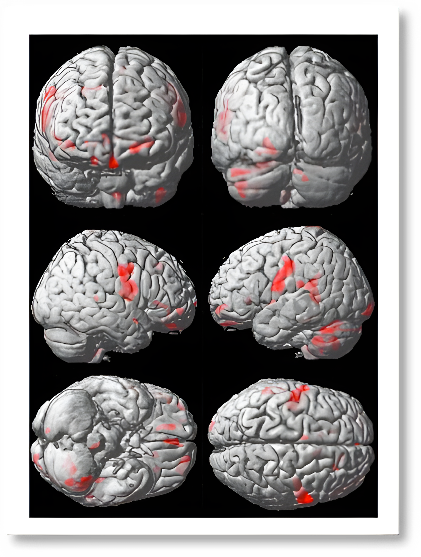
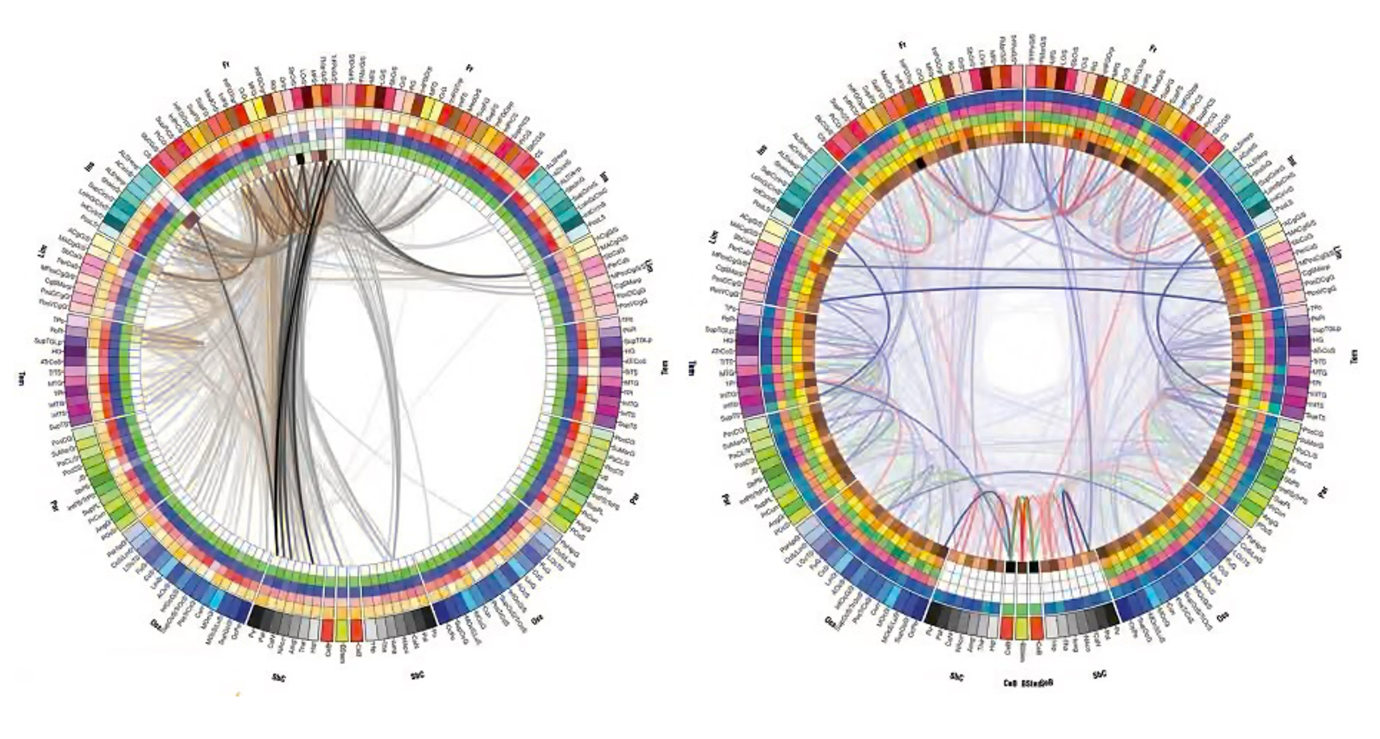
However, according to scans of her brain seen above, no significant damage was caused, so this effect must have been generated due to stress, which raises dopamine levels in the brain. Dopamine modulates prefrontal cortex inputs to the amygdala, and then to the hippocampus.
A bit about stress- high-stress levels disrupt the formation of memories as the amygdala is not regulated. Stress causes structural changes in the hippocampus via glutamate blockages which causes long-term depression (a decrease in the ability to release neurotransmitters) of synapses in the hippocampus, as well as harming their long-term potentiation ability which could explain why the patient showed changes to her memory capabilities. She ultimately started speaking in a forein accent to one she had never spoke-which is quite peculiar
Personality changes post trauma - Phineas Gage
This is the most famous case of brain trauma in history:
In 1984, Phineas Gage was working on a railway, when a three-foot-seven-inch, thirteen-and-a-half-pound tamping iron fired through his skull, entering beneath his left eye, and severing his optic nerve as seen in the render below. The parts of the brain that were destroyed are argued, but it is mainly the central area of the left hemisphere, but not too rear as the language and posterior motor areas were operating as normal which was concluded as there were no disruptions to movement or speech. This is still heavily debated, however, as there's very little evidence apart from reports of the severity of the damage to his personality.
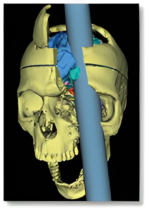
Computer mapping in 2012 allowed neurologists to observe the destruction of 11% of the white matter in the frontal lobe (what allows for the exchange of information in the brain- it is made up of large collections of axons) and 4% of the cerebral cortex (which is responsible for traits including intelligence and personality).
This injury is said to be the starting point of neuroscience and the most notable injury in neuroscience history. It provided great advancements in the mapping and understanding of the brain, such as the discovery (in 2022) that the brain can selectively use non-injured areas of the brain to help perform the functions lost during the trauma.
The symptoms of his trauma were dramatic changes in emotion, observed as he became aggressive and a heavy drinker, compared to being “energetic and motivated” prior to the trauma. This case is particularly interesting to me, and all neurologists, as Gage was able to survive and heal relatively quickly despite the severity of the injury, exposing the brain's resilience towards damage.
This ability to recover is what causes the changes in him (to some degree) as the brain reestablished neural pathways to resume as high a level of operation as possible, ridiculously allowing Gage to live for nearly 12 years after the event.
How symptoms of trauma are treated:
Mesenchymal Stem Cell trial for haemorrhages
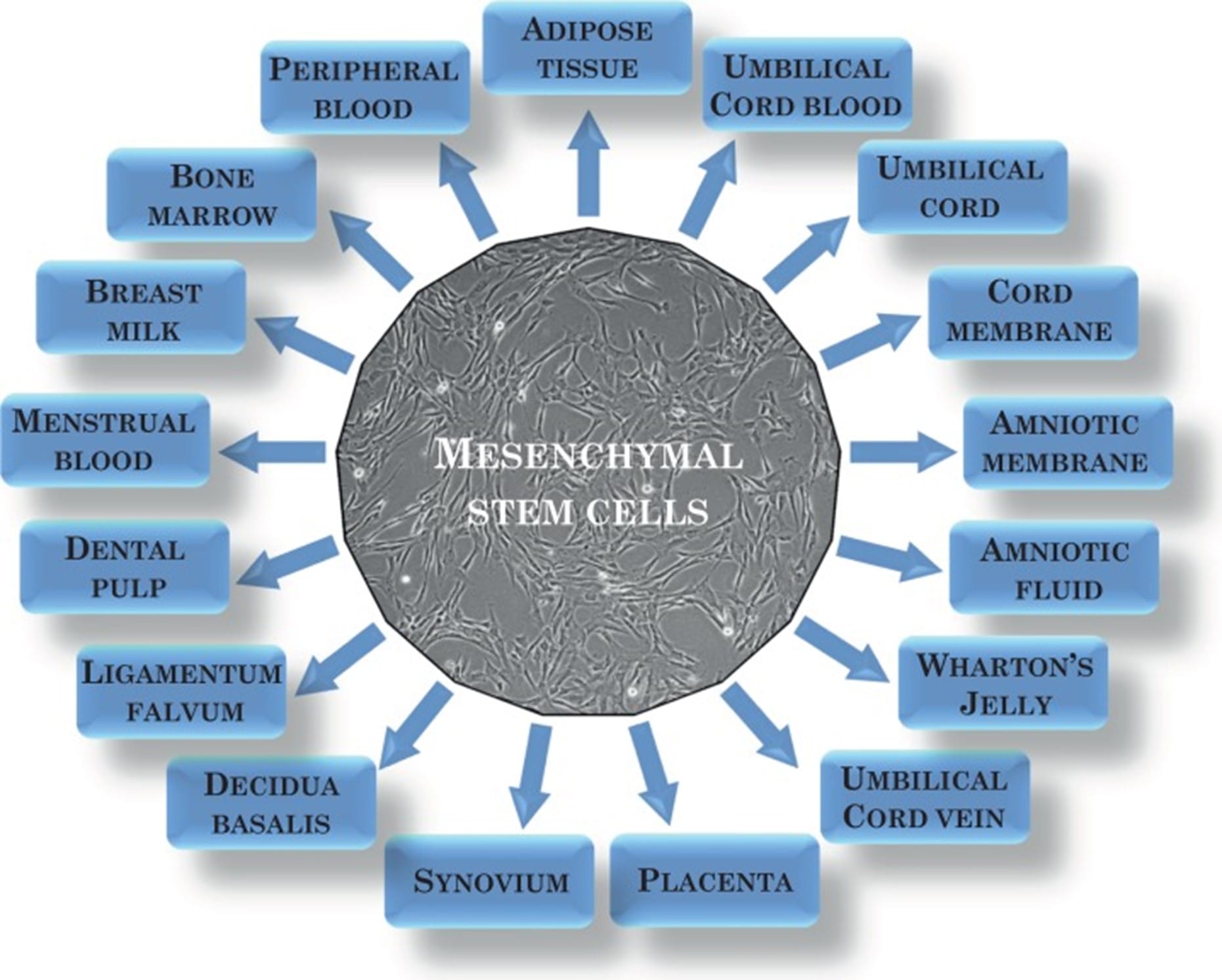
Mesenchymal Stem Cells are a type of non-specialised cells which reproduce quickly and are able to differentiate, first discovered by Friedenstein et al in the 1960s. They can be sourced from bone marrow, adipose tissue and birth-associated tissue like the placenta and umbilical cord (all sources can be seen in the image), with those from the umbilical cord appearing to have the most promise in neuroscience due to the ease to culture, isolate and some further unique properties.
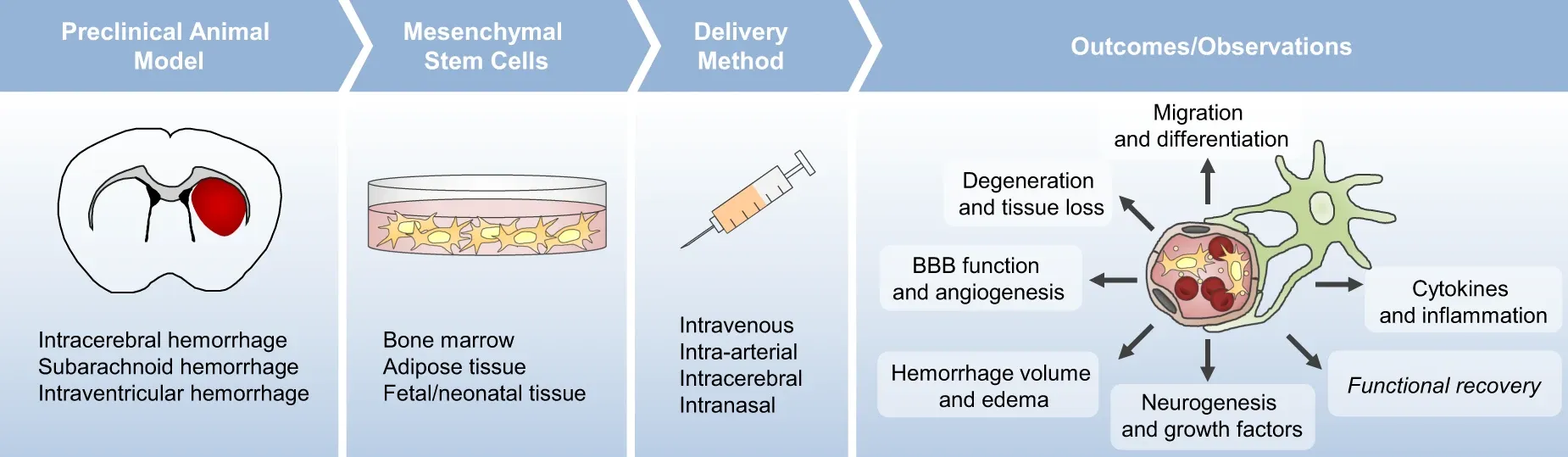
The concept of how these work in Neurotherapy is that they are inserted into the area of trauma (either by injection or delivery via robotics) where they are able to differentiate into neurons and reactivate the neural pathways that were previously depressed. This is still in heavy testing (with the Mayo Clinic being one of the leading institutes in this type of therapy) as it requires the body to not reject the cells.
Shown below is the process of using mesenchymal stem cells for a haemorrhagic stroke, done intravenously (via injection)
Music-based therapy
Music-based therapy has the most potential in the field of recovering after significant or minor brain injuries as music can activate more of the brain than any other stimulus, therefore reactivating and re-strengthening neural pathways and synapses to restore brain activity. This process is due to neuroplasticity, where the brain can reorganise pathways in response to changes in the environment.
Music is particularly useful for diagnosing what state of consciousness an individual is in, whilst allowing improving the quality of those experiencing significant brain trauma.
It has been shown to particularly develop muscular and fine motor coordination as well as sensory responsiveness, being helpful to those with damage to the cerebellum and motor cortex. It stimulates positive moods, and memories and allows expression despite a lack of ability to communicate verbally.
Conclusion
In conclusion, neurology is a fascinating subbranch of medicine which varies from research to surgery and treatment.
It truly is a subject that will transform the near future such as developments in robotics and AI such as the research by companies such as Neuralink (by Elon Musk) which aims to allow paralysed individuals to control computers via their brain. Other developments include current attempts to synthesise human-level consciousness for an increased capacity of AI.
Regardless of this, however, the human brain is a very complex mechanism and feat of evolution allowing for conscious thought, emotion and the storage of memories and is what has allowed humans to progress to this point in civilisation and continue to develop exponentially in all sectors of life.
Thank you for reading this article, I have found it to be an interesting study and I hope you have enjoyed it too.

![Image via [Adobe Stock]](/content/images/size/w600/2026/03/IMG_0874.jpeg)
